Plant tissue types: dermal, vascular, and ground tissue
Plant tissue is often described as having three main systems: dermal tissue forms the outer covering of roots, stems, and leaves; vascular tissue carries water and sugars; and ground tissue makes up much of the interior and handles the rest. Mix them up, and root, stem, and leaf structures start to blur. This guide shows how to spot each tissue in roots, stems, and leaves.
What are the three main plant tissue types?
Plant tissue is usually grouped into dermal, vascular, and ground tissue. Those three systems divide labor cleanly: protection at the surface, transport inside, and bulk functions through the rest of the organ. That split makes plant structure easier to read in cross-section, especially when the tissue pattern changes by organ.
Basic plant anatomy pages often name the same three groups. That matches the way I read a stem or leaf slice under a microscope: an outer layer, transport strands, and everything filling the spaces between and around them. In the plant structure chapter, tissues are taught as part of body organization, not as isolated cell names.
Why plants use separate tissue systems
One tissue cannot do every job well. Surface cells have to block water loss and take damage first. Transport cells need open pathways and strong walls. Fill tissue needs room for photosynthesis, storage, and support. Splitting those jobs lets each tissue specialize.
That is why the same organ can contain all three systems at once. A leaf has an outer dermal layer, vascular bundles through the blade, and ground tissue between them. A stem and root do the same, but the layout changes.
How do you spot dermal tissue?
Dermal tissue is the outer protective tissue. In young organs it often appears as the epidermis, a single outer cell layer that can help reduce water loss and defend the plant. In leaves, the epidermis often carries a cuticle and stomata, which makes it easy to find in cross-section.
Epidermis, cuticle, and stomata
The epidermis is the first line of defense. It sits on the outside of young roots, stems, and leaves. In leaves, a waxy cuticle often covers it and stomata interrupt it for gas exchange. Those openings matter because they also create the main route for water vapor loss.
In roots, the epidermis is usually thin and focused on absorption. In stems and leaves, the outer layer is more about protection and control of water loss. That shift is one reason dermal tissue looks different from organ to organ even though it keeps the same basic job.
What goes wrong when the outer layer is damaged
If the epidermis is missing or damaged, the plant loses water faster and becomes easier to infect. Leaves can wilt sooner. Young stems can dry out. Roots can take up less water if the outer surface is injured. Dermal tissue is not decorative; it is the plant’s front barrier.
What does vascular tissue do in plants?
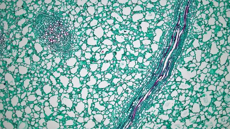
Vascular tissue is the transport system. It includes xylem and phloem, which are often arranged together in vascular bundles in stems and leaves. Xylem carries water and dissolved minerals, often upward, while phloem moves sugars and other organic compounds from source to sink. It also adds support.
Xylem: water, minerals, and support
Xylem moves water and dissolved minerals from roots to the rest of the plant. The main conducting cells are vessels and tracheids. Their walls are strengthened, so xylem helps with structural support too. When someone sees a stiff vein or a hard strand, xylem is often part of it.
Damage to xylem is serious because the transport path breaks. Water cannot move properly to leaves, so tissues above the injury can wilt or dry out. In woody plants, a blocked xylem pathway can also weaken the stem because transport and support are linked.
Phloem: sugar transport from source to sink
Phloem moves sugars and other organic compounds from source tissues, usually photosynthetic leaves, to sink tissues that use or store them. Its conducting cells are sieve tube elements, and companion cells help keep them working. The direction is about source and sink, not a simple upward-only route.
Phloem and xylem often sit together in the same vascular bundle. That pairing lets water and food move through the same organ without mixing jobs. In stems and leaves, vascular bundles are the easiest place to find both tissues at once.
What counts as ground tissue?
Ground tissue fills most of the plant body. It includes parenchyma, collenchyma, and sclerenchyma, and it is found in areas such as the cortex, pith, and mesophyll. This system handles photosynthesis, storage, and support, so it forms the background tissue between the dermal layer and the vascular system. (en.wikipedia.org)
Parenchyma, collenchyma, and sclerenchyma cells
Parenchyma cells are living, thin-walled ground tissue cells. They are common in storage and photosynthesis and appear in the cortex, pith, and mesophyll. If a tissue looks soft, flexible, and full of living cells, parenchyma is a strong possibility.
Collenchyma is living support tissue with unevenly thickened primary walls. It gives flexible support in growing organs and often sits just beneath the epidermis in stems and petioles. Sclerenchyma is different: it has thick lignified secondary walls and is often dead at maturity. Fibers and sclereids belong here.
Cortex, pith, and mesophyll
The cortex is the ground tissue between the epidermis and the vascular cylinder in roots and stems. The pith is the central ground tissue region in many stems. Mesophyll is the ground tissue of leaves, where photosynthesis happens. Same system, different location, different look.
The LibreTexts plant tissue terms list epidermis, parenchyma, collenchyma, and sclerenchyma together, which fits the idea that tissues are part of plant structure and cell organization. Ground tissue is the broad category that holds those cell types together.
How are roots, stems, and leaves different in cross-section?
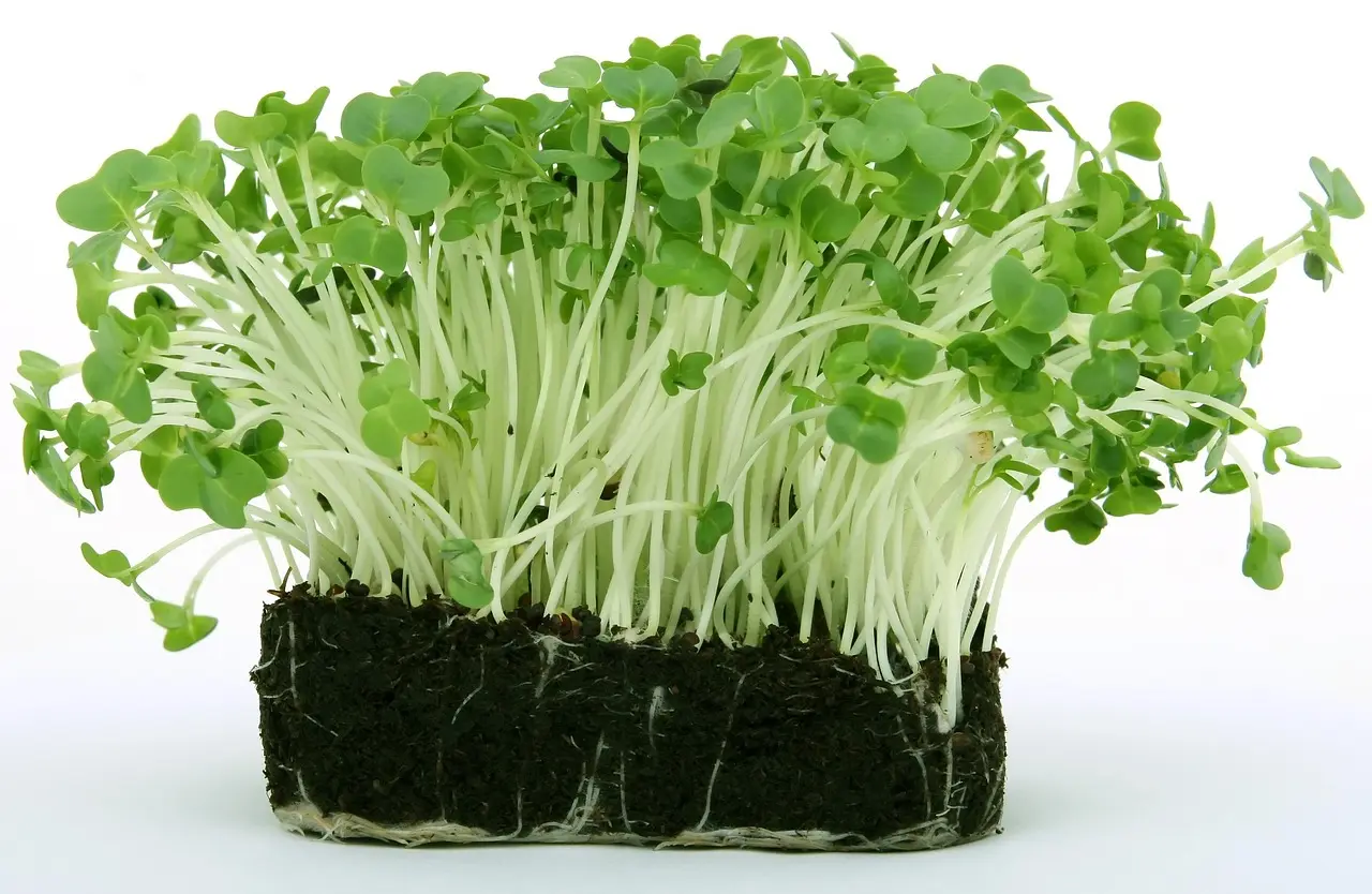
Roots, stems, and leaves all contain dermal, vascular, and ground tissue, but they arrange them differently. Roots often keep vascular tissue near the center. Stems often place vascular bundles in a ring or scattered pattern. Leaves spread vascular bundles through the blade, with mesophyll around them.
Root tissue layout
A root cross-section usually shows an outer epidermis, a broad cortex of ground tissue, and a central vascular region. The pattern is built for absorption and transport. The root is not trying to photosynthesize, so its ground tissue is not arranged like leaf mesophyll.
Roots are the easiest place to confuse cortex with vascular tissue if the slice is thin or damaged. The cortex is ground tissue, not transport tissue. The vascular tissue is the inner conducting core.
Stem tissue layout
A stem often has epidermis on the outside, collenchyma or other ground tissue beneath it, and vascular bundles through the interior. Those bundles contain xylem and phloem together. The rest of the stem may be pith inside and cortex near the edge, depending on the plant group.
In stems, collenchyma is often just under the epidermis, which gives flexible support where growth is active. Sclerenchyma may also appear near bundles or as tougher support bands. That is why a stem slice can look layered instead of fully mixed.
Leaf tissue layout
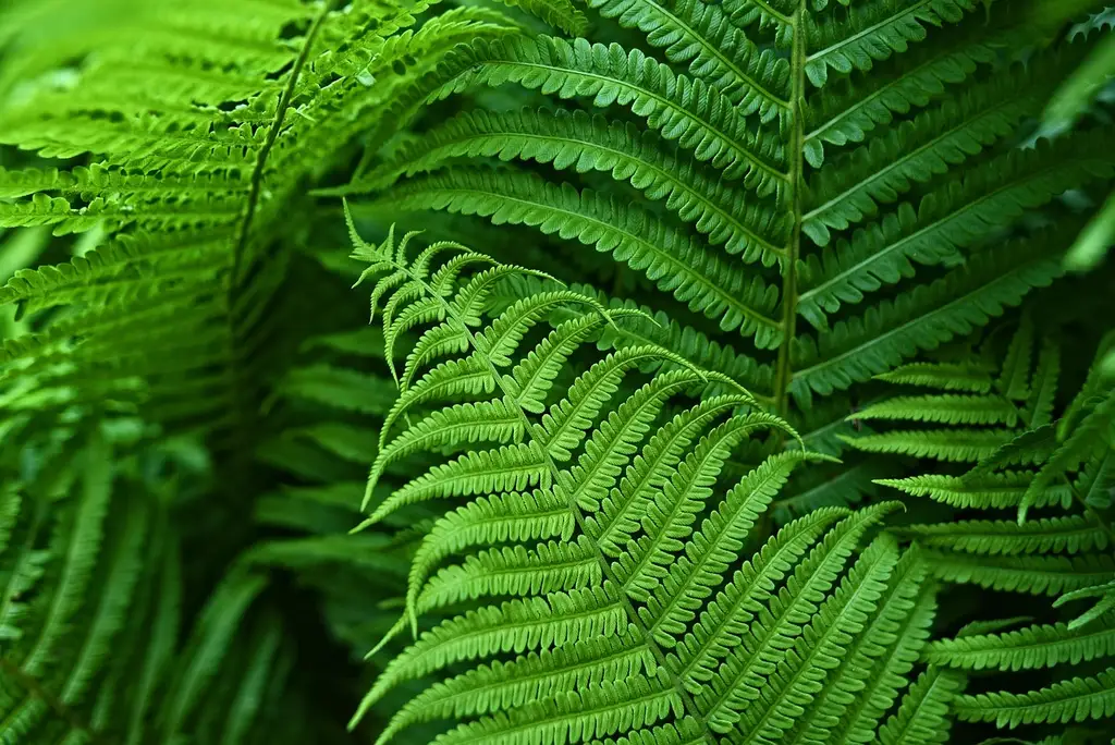
A leaf usually has epidermis on both surfaces, vascular bundles in the veins, and mesophyll as the main ground tissue between them. The mesophyll is often packed with parenchyma cells for photosynthesis. Stomata in the epidermis help control gas exchange.
Leaves are the clearest example of tissue jobs matching position. The outside protects, the veins transport, and the middle does most of the food making. Once that pattern is familiar, a leaf cross-section becomes easier to read at a glance.
Use the comparison table to identify each tissue system
This table is the fastest way to sort a cross-section. Read down the organ column, then compare the job, the visible clue, and the common mistake. If the sample has an outer layer, transport strands, and a filling tissue, the three systems are all probably present.
| Tissue system | Roots | Stems | Leaves | How to identify in cross-section | Common mistakes |
|---|---|---|---|---|---|
| Dermal tissue | Epidermis on the outside | Epidermis on the outside, often with a cuticle in young parts | Upper and lower epidermis, often with stomata | Look for the outermost cell layer that forms the boundary | Do not call cortex or pith “outer tissue”; those are ground tissue |
| Vascular tissue | Central conducting region | Vascular bundles arranged in a ring or scattered pattern | Veins and bundles through the blade | Find xylem and phloem together as a bundle or core | Do not confuse veins with the whole leaf tissue; veins are vascular only |
| Ground tissue | Cortex, with central support tissues depending on the root | Cortex and pith, plus support tissues around bundles | Mesophyll between the epidermis layers | Look for the tissue that fills the spaces around transport tissue | Do not mistake parenchyma-filled regions for vascular tissue just because they sit near veins |
Reading the table by organ and job
Start with the organ, then ask what problem the tissue solves. In roots, the key job is uptake. In stems, the key jobs are support and transport. In leaves, the key job is photosynthesis with gas exchange. The tissue layout changes to fit those jobs.
A practical memory aid
Use this sequence: outside, lines, filling. The outside is dermal tissue. The lines are vascular tissue. The filling is ground tissue. If a cross-section has more than one of those zones, the organ is doing more than one job at once, which is normal.
How do the tissue systems work together?
The three systems work as a set. Dermal tissue protects and limits water loss. Vascular tissue moves water, minerals, and sugars. Ground tissue stores, photosynthesizes, and supports. A plant needs all three because each one depends on the others for normal function and survival.
What plants lose when one tissue is damaged
If dermal tissue is damaged, the plant loses water faster and becomes more exposed to pathogens. If xylem is damaged, water supply fails and upper tissues can collapse. If phloem is damaged, sugars cannot reach growing or storage tissues well. The effect depends on which system fails first.
That is also why vascular bundles matter so much. They are not just tubes. They are transport plus support units. In stems and leaves, their arrangement can change by plant group, but the paired xylem-and-phloem pattern stays the same.
Frequently asked questions
What are the three main plant tissue types?
The three main plant tissue types are dermal tissue, vascular tissue, and ground tissue. Dermal tissue forms the outer skin and protects the plant surface. Vascular tissue carries water and sugars. Ground tissue fills most of the plant body and handles storage, photosynthesis, and support.
What does dermal tissue do in plants?
Dermal tissue forms the plant’s outer protective layer. In young organs, it usually includes the epidermis. In leaves, it can also include the cuticle and stomata. Its main jobs are protection, water-loss control, and defending the plant from damage and infection.
What is the difference between xylem and phloem?
Xylem moves water and dissolved minerals, mostly upward from roots toward shoots, and it also adds support. Phloem moves sugars and other organic compounds from source to sink. They work together in vascular bundles, but they carry different materials and solve different transport problems.
What counts as ground tissue in a plant?
Ground tissue includes parenchyma, collenchyma, and sclerenchyma. It makes up most of the plant body and is found in the cortex, pith, and mesophyll. Its jobs include photosynthesis, storage, and support, so it fills the space between dermal and vascular tissue.
How are epidermis, cortex, and vascular bundles different?
The epidermis is dermal tissue and forms the outer cell layer. The cortex is ground tissue found beneath the epidermis in many roots and stems. A vascular bundle is a package of xylem and phloem, usually in stems and leaves, and it handles transport.
Why do plants need multiple tissue types instead of one?
Plants need multiple tissue types because different jobs require different cell designs. Surface protection, long-distance transport, storage, photosynthesis, and support cannot all be done well by one uniform tissue. Dividing labor lets each system specialize, which makes the whole plant more efficient and easier to identify.